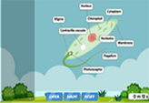
Amoeba cell diagram
An amoeba is a microscopic organism with a wacky, constantly-changing shape. It can be dangerous once it infects a human host, although by observing proper safety guidelines at your lab, it’ll be perfectly fine to observe and experiment on these microbes.
Amoebae are a type of eukaryotic organism made up of only a single cell. They look like living jelly. Unlike other cells, amoebae don’t have a protective cell wall, although they can “harden” against poor conditions, such as summer’s heat or winter’s cold, to survive for a while longer.
Their defining feature is the pseudopod; the amoeba is able to project a part of its body outward to move and catch food. Several pseudopodia can be created at any time.
Amoebae also have cytoplasm, the jelly-like substance in which all of its other body parts are contained. Within the cytoplasm lies its nucleus for reproduction, as well as a bunch of mitochondria for converting globules of food into energy.
The organism also has sacs called vacuoles which help in storing nutrients and expelling waste. These waste products float around the cytoplasm in the form of crystals.
Find out more about how amoebae live, behave, and feed with our own amoeba cell diagram.

